 Are you a Glaucoma Suspect? Learn about the Mystery behind Glaucoma
Glaucoma is a group of eye diseases that can lead to permanent vision loss when left untreated. A patient with glaucoma often won’t notice any symptoms, yet the eye disease will secretly & slowly damage the optic nerve due to an imbalance of eye pressure. Since the optic nerve is solely responsible for sending signals to the brain, a damaged optic nerve will lead to permanent vision loss.
The nature of Glaucoma’s subtle changes causes optometrists to take extra care when evaluating patients who potentially may have glaucoma. In the industry, patients that display any signs of Glaucoma are labeled a Glaucoma Suspect.
What is Glaucoma?
The cornea or eye surface has a layer of fluid to create a protective barrier against foreign bodies. The fluid naturally drains out of the eye to allow new fluid to enter. When the fluid fails to drain out of the eye at the normal rate, the inside of the eye increases in pressure due to the excess fluid. This imbalance of pressure will lead to optic nerve damage.
- Primary Open Angle Glaucoma (POAG) is the form of glaucoma when the eye’s drainage canals are blocked.
- Angle Closure Glaucoma is a result of a narrow or closed off drainage canal. Due to the severity of this form of glaucoma, eye pressure can rise very quickly and can be triggered by pupil dilation. This form of glaucoma often bears symptoms of eye pain, headaches, blurred vision, and nausea. Angle Closure Glaucoma is an eye emergency and requires immediate attention from an eye doctor. (This form of glaucoma is less common than POAG.)
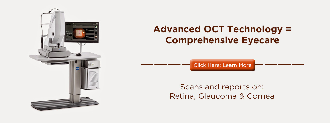
- Normal Tension Glaucoma (Low-Pressure glaucoma) is a difficult form of glaucoma for an optometrist to detect as the eye pressure remains near-normal. Through an OCT scan or other digital retinal imaging, an eye doctor can examine the optic nerve to determine any eye damage. While the eye pressure isn’t high, reducing eye pressure for this type of glaucoma will still treat glaucoma.
- Secondary Glaucomas result from eye complications, such as cataracts, diabetes, post-eye surgery, tumors, and even eye trauma. Medication or surgery will treat this form of glaucoma.
Depending on the form of glaucoma, the standard puff test can display signs glaucoma early on. Diagnosis of glaucoma, however, based on eye pressure alone isn’t enough to detect Glaucoma until after the disease has progressed. Glaucoma has been known in the industry as the “sneak thief of sight” since the actual disease carries no symptoms until the disease has already progressed to a severe level. This is why optometrists utilize digital retinal imaging for a more precise diagnosis.
Optometrists label certain patients as Glaucoma Suspects
Depending on your eye exam with an optometrist, the state of your eye health may classify you as a glaucoma suspect.
Here are some indicators that could be used:
- Elevated intraocular pressure
- Over the age of 40
- Family history
- Prior eye trauma
- The health and appearance of the optic nerve
- Eye inflammation
- Race, more common by African Americans
While being labeled a glaucoma suspect has no bearing whether you have glaucoma or not, your eye doctor may recommend more digital testing, such as an OCT scan to review the health of the optic nerve.
If you fall under some of the indicators above, speak to one of our eye doctors. We’ll review your medical history and ways to ensure your eyes remain healthy and how to prevent any vision damage from glaucoma.

