 It is important to have regular comprehensive eye examinations. It is not just about checking your vision but there are so many more problems that we need to look for to help protect your eyes and health.
During a complete eye exam, we not only determine your prescription for eyeglasses or contact lenses but will also check your eyes for eye diseases, assess how your eyes work together as a team and evaluate your eyes as an indicator of your overall health.
We perform many tests and procedures to examine and evaluate the health of your eyes and the quality of your vision. These tests range from simple ones, like having you read an eye chart, to complex tests, such as using a high-powered lens to examine the health of the tissues inside of your eyes.
What Vision Problems Do We Look For:
Everybody knows about using an eye chart to measure vision and the instrument we use to determine an eyeglass prescription. But there are many other tests we do to find any eye and systemic medical problems. Some of the instruments we use and testing that we perform are:
1. Visual field testing:
We use a Humphrey 750i which is a sophisticated instrument for measuring central and peripheral vision. For example, we have found brain tumors, glaucoma damage, and peripheral vision loss from strokes with our Humphrey 750i.
2. Measuring Eye Pressure:
This is sometimes referred to as “the glaucoma test”. However, eye pressure alone can not tell you if a person has glaucoma. If the pressure is elevated, we have to do additional testing such as OCT scans, visual fields, and optic nerve study to see if the person truly has glaucoma.
Importantly, people do not realize they have glaucoma until the disease is advanced and there is significant permanent eye damage.
This is a very important reason to have yearly eye examinations.
 3. OCT Analysis.
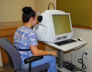
We use a state-of-the art Cirrus 5000 OCT to visualize the retinal and macular layers to look for eye diseases. We also use it to help diagnose glaucoma and to help verify that the treatment for glaucoma is controlling the disease properly.
The photo to the right shows how an OCT scan is performed on a patient.
The image below from an OCT scan shows a healthy, normal macula.
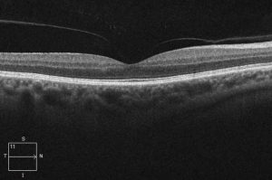
The image below shows a macular detachment which is a serious condition.

4. Retinal Photography:
This is another very important tool to look for eye and systemic medical problems. There are so many problems that can be detected via retinal photography.
For example, we have seen many cases of:
- early macular degeneration even before there are symptoms
- retinal bleeding in diabetes
- some types of brain tumors
- cholesterol plaques that indicate clogged carotid arteries that tell us the person is at risk of stroke
- and countless other conditions.
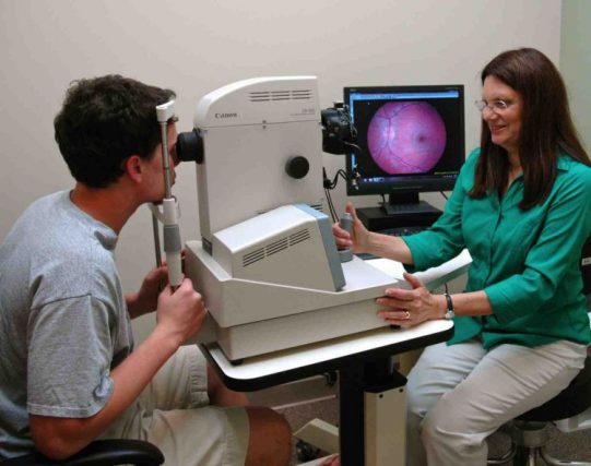
The photographs below show Dr. Arcolano taking a retinal photograph.
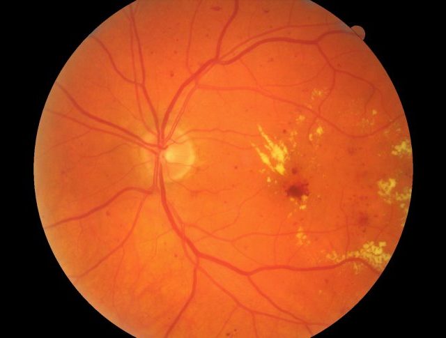
Retinal Bleeding and Edema from Diabetes
We referred this individual to a retinal specialist for treatment.
Retinal Imaging Can Detect Life Threatening Conditions
These photographs below are from a 22 year old person who came for a routine examination. They had been having severe headaches for several months.
We discovered their optic nerve was swollen. This is a serious, potentially life-threatening condition, which is caused by raised fluid pressure in the brain.
We sent them directly to the ER. The next photo is the same person a few months later after treatment. The problem was resolved and the headaches were gone.
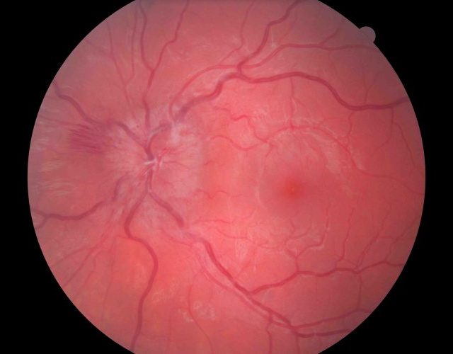
Swollen Optic Nerve of a 22 year old.
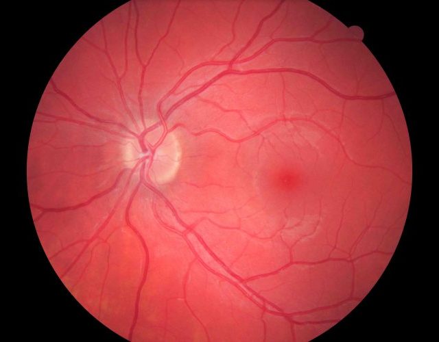
The Optic Nerve Back to Full Health
5. Biomicroscopy:
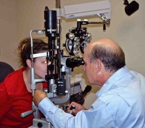
This is a high powered microscope we use to study the front half of the eye. We find:
- cataracts
- corneal problems
- lid problems
- dry eye damage
- many other conditions with this testing.
For example, we have found a number of people with basal cell cancer on their eyelids.
The photo below shows Dr. Vaughan performing biomicroscopy.
6. Other Testing
There are other testing and procedures that are done as needed, such as:
- corneal topography
- dry eye testing
- gonioscopy
- auto-refraction
- fluorescein testing for corneal problems.
When Should You Have an Eye Examination?
Children: Children have less eye disease than adults but they frequently have vision problems. We see many children with eye problems and we find that these children rarely complain to anybody about their vision. The most common problems are amblyopia ( “lazy eye”), and large amounts of farsightedness and/or astigmatism.
There can be permanent vision loss if this is not discovered in time and treated with eyeglasses and sometimes eye patching. For example, treatment of vision loss from amblyopia is very difficult in an eight year old but easy in a four year old. A particularly serious red flag of problems is when a child has an eye that turns in constantly or even occasionally. Even if there are no signs of problems we recommend a first eye examination at age four and then yearly thereafter. We recommend bringing in a child of any age for a pediatric eye exam if their eyes cross or if you see an indication of any vision problems.
Common risk factors for vision problems include:
- premature birth
- developmental delays
- turned or crossed eyes
- a family history of eye disease
- history of eye injury
- other physical illness or disease
Adults: We recommend yearly examinations for all adults, regardless of their vision. Some adults need to be seen more often, for example, those with retinal bleeding from diabetes, or those undergoing glaucoma treatment. We also encourage you to call our office if you have any specific concerns about your eyes or vision or if you see any unusual changes. We will see you promptly if you have any symptoms of serious problems.
Call us on (910) 423-8600 to schedule your eye exam now or come visit us. Both Dr. William Vaughan, O.D. and Dr. Laurie Arcolano, O.D. have been providing eye care in Fayetteville, NC for many years – Village Eye Care is conveniently located on Village Drive two blocks from the Cape Fear Hospital.

