Eye Exams for Adults Age 65 & Over
Why are geriatric eye exams so important?
As you age, your risk of eye disease and vision problems rises. If you notice any unusual visual symptoms, it’s wise to have an eye exam performed as soon as possible by an eye care professional. Additionally, many common ocular conditions associated with aging can linger for a long time without symptoms, yet still causing damage. This underscores the importance of routine geriatric eye exams, which can detect signs of disease before you do! The earlier an eye disease is diagnosed, the earlier treatment can begin. This goes a long way towards preventing blindness, partial vision loss and complications.
When should older adults have comprehensive eye exams?
According to the American Academy of Ophthalmology, adults age 65 and older should have their eyes evaluated thoroughly every year or two. Many common eye diseases, such as glaucoma and diabetic retinopathy, are silent at the beginning and progress very slowly. Experts have estimated that approximately half of the American senior population has glaucoma and doesn’t know it.
Certain health conditions, such as hypertension and diabetes, point to a need for more frequent eye exams. Other risk factors include a serious past eye injury, personal family history of eye disease, being African American (has a higher risk of glaucoma) and specific medications. Your eye doctor should be consulted regarding how often to schedule a geriatric eye exam.
What are some eye symptoms that need an eye exam immediately?
If you, or a senior in your care, experience any of the following symptoms, an eye examination should be reserved as soon as possible. These may be signs that a serious condition has developed.
- Seeing flashes of light or new floaters
- Excessive tearing or discharge
- Vision loss, including blind spots or a dark spot in the center field of vision
- A change in the eye’s appearance, such as crossed-eyes, bumps, bulging or swelling
- Double vision
- Seeing halos
- Blurred or cloudy vision
- Straight lines appearing wavy
- Eye pain or fatigue
- Inability to close eyelid
- Burning or itchiness
What tests are performed in a geriatric eye exam?
Your eye doctor will pay special attention to inspect for signs of age-related ocular diseases, such as glaucoma, cataracts, macular degeneration and diabetic retinopathy. The procedures that will be done are not painful.
Here are the basic steps of what will happen in an eye exam for seniors:
- An evaluation of the outside of your eyes
- Visual acuity testing: you will need to read a standard eye chart of letters, placed at 20 feet away, first covering one eye and then the other eye. This will determine your vision prescription or need for eyeglasses.
- Refraction assessment: by checking how light waves are bent when they pass through your cornea and lens, an eye doctor will be able to diagnose nearsightedness, farsightedness, presbyopia and astigmatism. This procedure is done by having you look through a series of lenses to provide feedback about which lens is clearest.
- Visual field testing: peripheral (side) vision is checked using a computer that flashes lights on either side of you
- Color Vision: you’ll be tested for color blindness by looking at images formed by multicolored dots and stating what you can see
- Eye mobility: eye muscle movement will be assessed by having you move your eyes in different directions
- Ocular health: your eye doctor will use a slit lamp, which is a magnifying tool, to inspect all the parts of your eyes, such as the iris, cornea, retina, macula and optic nerve. Dilating eye drops may be inserted to enable a comprehensive examination of the back of your eyes
- Glaucoma testing: a specialized test, using tonometry, is performed to measure the inner fluid pressure of your eye
What is a diagnosis of Low Vision?
Low vision refers to a loss of sharp eyesight that cannot be corrected fully by eyewear. Among other age-related changes that occur to your visual system, glaucoma, diabetic retinopathy and macular degeneration can all lead to low vision. Poor visual acuity is not the only indicator of low vision, and other skills, such as limited peripheral vision and weak depth perception can also limit daily functioning.
Low-vision specialists can help maximize remaining eyesight through a variety of strategies and rehabilitation options. In this way, your independence and quality of life can be maintained. Many people with low vision can still work and live well and safely. Low vision devices are also very helpful, including eyeglass-mounted, handheld and stand magnifiers (mounted on eyeglasses or a headband), miniature telescopes and video magnification systems.
Nowadays, many seniors enjoy an active, healthy lifestyle throughout their golden years. Geriatric eye exams contribute significantly to this, as they allow eye diseases and low vision problems to be detected and treated as early as possible, before they severely damage your eyesight.
OCT & Optos Testing
OCT – Optical Coherence Tomography
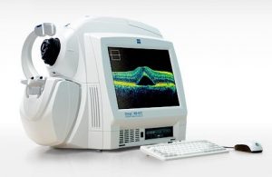
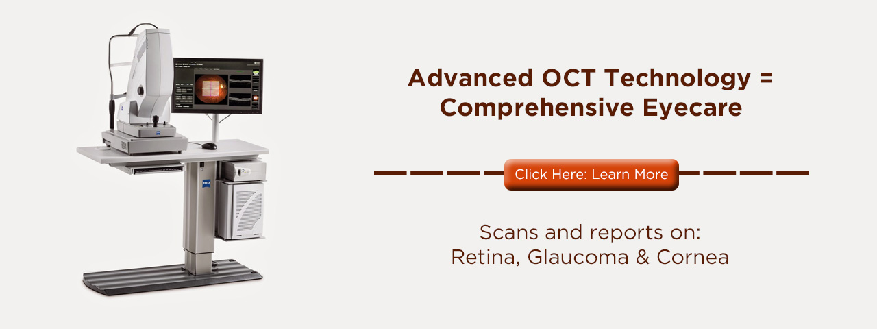
Optical Coherence Tomography (OCT) is a scanning technique that allows an optometrist to look deeper into the patients’ eyes that would be possible during a regular eye exam, for the purpose of diagnosing eye diseases. Although many optometric practices do not yet provide OCT Mississauga Vision Centre is proud to offer the latest technology in eye care.
 Zeiss, world-renowned for its cutting-edge lens technology, produces this high definition 3-dimensional scanner. With fast-acting retinal tracking, precise macular thickness analyses, and detailed layer maps, this advanced OCT scanner allows us to pick up early warning signs of developing macular degeneration, and monitor confirmed cases for progression, allowing earlier intervention which is associated with better vision loss prevention.
Does It Hurt?
OCT scanning is non-invasive: it doesn’t even touch the eye, and the results are instantaneous. You just look into the eyepiece, and the OCT produces a 3D image of the layers of the retina. The way a dental x-ray yields an image of the root of the tooth, OCT helps to visualize the retina, which can be compared to the root of the eye. Changes to the sensitive tissue of the retina can indicate the development of serious eye diseases and conditions. Treating them early can save eyesight. That’s why our eye doctors offer this advanced diagnostic technology at our Village Eye Care office.
Optos Retinal Imaging
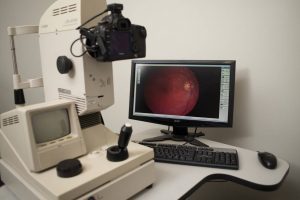
 Our doctors are committed to providing you with the highest standards of eye care available and that is why our doctors strongly recommend that you have an Optomap Retinal Exam as part of your eye exam. The Optomap Retinal Exam provides important information about your health, without the need for uncomfortable pupil dilation. With the Optomap Retinal Examination, the majority of the retina is visible in one single image. Obtaining images is comfortable, quick and very simple.
 By looking into the Optomap eyepiece the digital image of your retina is captured in less than a second. A small flash indicates the image has been taken. Nothing touches your eye during the process. Your Optomap Retinal Image is then immediately displayed for your eye specialist to review with you. Best of all it does not require dilating drops which typically cause blurred vision and sensitivity to light for hours. Click to watch a video on the benefits of the Optomap.


